In today's time, the coronavirus pandemic has caused havoc in the world. This is evidenced in the upsurge of research that is being conducted by both public and private sector laboratories. However, there is a lot of curiosity from the public and individuals are asking the question as to how COVID-19 is being investigated in the laboratories? Keeping this in mind, we have tried to provide information in this article about the detection of COVID-19, thus we hope this information will be helpful to understand the detection of the COVID-19 disease using polymerase chain reaction (PCR). Therefore, we will first know what are PCR and the variations thereof between PCR, RT-PCR, qPCR, and RT-qPCR and how they work. We will further explain how corona virus is detected through RT-PCR.
Key Words: COVID-19, PCR, RT PCR, RT qPCR
PCR is a laboratory (in-vitro) technique that is employed for amplifying a targeted portion of DNA. This technique is a cell-free method utilized to synthesize several identical copies of a gene or DNA of interest. Thus, it is a very essential tool in a molecular biology laboratory synonymous to a photocopier as a basic requirement in an office. First, we will outline the requirements for a standard PCR reaction – the following will be needed: DNA polymerase enzyme, magnesium chloride (MgCl2), nucleotides, primers, nuclease free water, target DNA template to be amplified and a PCR machine. The PCR reaction mechanism is simple and starts with an initial Denaturation where the double-stranded DNA is heated to separate the strands (1st step) this allows for the primers to attach to the now single stranded DNA (2nd step). After the primers align to the DNA strand, the DNA polymerase enzyme extends the primers (3rd step) which results into two identical copies of the original DNA template. These three steps are repeated over a series of temperatures and times a single complete process of the three combined is known as an amplification cycle. It is important to optimize every step of the cycle to the DNA template and the set of primers being used. The cycles can be repeated as many times as needed, however, on average a standard PCR is run for around 30 to 40 times after which the amplified product is then analyzed. The polymerase chain reaction is used to amplify a targeted DNA of interest for downstream experimental purposes; and it has also found immense use in detection of pathogenic DNA as well as other applications in genetic testing. PCR is a very sensitive technique as tiny volumes of a single reaction are required, as such it is recommended that a master mix is prepared if there are many reactions. The master mix is thoroughly mixed and split according to the number of reactions ensuring that each reaction contains an equal amount of the reaction mixtures outlined earlier. A number of companies are now available which supply ready to use mixes except for primer and the target DNA. It should be mentioned that the Guanine and Cytosine-rich regions, commonly referred to as the GC regions, may be a constraint in conducting a normal PCR as these regions tend to be more stable than those with lower GC content. Besides, GC-rich regions are prone to making secondary structures such as hairpin loops and are difficult to fully separate during the Denaturation part, thus the DNA polymerase enzyme may be hindered from synthesizing new DNA strands properly. In this regard, a higher Denaturation temperature helps to prevent this challenge and shorter annealing time avoids unspecific amplification. The addition of more reagents enhances amplification of GC-rich regions, and addition of DMSO, glycerol and betaine to the reaction helps to disrupt the secondary structures that may be caused by the GC interaction.
One drawback that may occur during PCR is unspecific amplification. Besides, most DNA polymerase enzymes that are used in molecular biology laboratories work best at 68 to 72oC. It is therefore suggested to choose the extension temperature in that range. The polymerase enzyme will nevertheless, be active to a lesser degree at even lower temperature. However, primers appear to bind to non-specific regions at temperatures that are much lower than the annealing temperature even if the reaction mixture is prepared on ice. In this regard, enzyme inhibitors may be used which dissociate from the DNA polymerases once the required temperature is reached. Since the inhibitor is an antibody that binds to the polymerase, it gets denatured during the initial denaturation temperature.
The reverse transcription PCR, commonly known as RT-PCR, uses RNA as a template. An additional step allows the detection and amplification of RNA after which it is reverse transcribed into its complimentary DNA (cDNA) by an enzyme called reverse transcriptase. The success of RT-PCR is dependent on the purity of the RNA. The initial step in conducting RT-PCR is synthesis of a DNA/RNA hybrid and the reverse transcriptase has an inbuilt rnase H function for degrading the RNA portion of the hybrid. The resultant single-stranded DNA molecule is then converted by a DNA-dependent polymerase activity of the reverse transcriptase into complimentary DNA. It is important to note that the amplification method will be affected by the potency of the first-strand reaction, but after this first step, the subsequent, the standard PCR procedure is utilized to amplify the cDNA. The approach to convert RNA into its complementary DNA by RT-PCR is advantageous in that RNA is single-stranded and extremely unstable which makes it difficult to work with, and second, it is commonly used as the first step in qPCR to quantify RNA transcripts in a biological sample.
Quantitative PCR (qPCR) is mainly used in differential gene expression, i.e. to monitor, characterize and quantify nucleic acids in biological samples. Briefly, in RT-qPCR, RNA is quantified by converting it to its complementary DNA as stated earlier followed by qPCR. Similar to the standard PCR, DNA is first amplified by the three consecutive steps - denaturation, annealing and extension. The only variation in qPCR is fluorescent labeling which allows the detection and collection of information as the reaction progresses. Fluorescent labeling provides several advantages as a range of options are chemistries are available. For instance, in dye-based qPCR (typically green), fluorescence permits to quantify the amplified DNA using a dsDNA biding dye and the fluorescence is measured whenever a double stranded binding occurs throughout the cycle and the degree of fluorescence is proportionate to the number of replicated DNA and it is quantified in real-time. The only disadvantage of dye-based systems is that one target only can be examined at a time to which the dye will bind within the sample. The other alternative to dye-based qPCR is probe-based in which several targets can be detected at the same time, however, this requires designing of target-specific probes and proper optimization in addition to primers. Many probe designs are available, but commonly used probes are hydrolysis based probes which comprise a fluorophore and quencher. The fluorescent resonance energy transfer (FRET) stops the fluorophore’s emission through the quencher and in turn makes the probe stay intact. The probe, however, is hydrolyzed throughout the PCR reaction as the primer extension occurs resulting into amplification of sequences that get bound to it. The probe has a cleavage part which separates the fluorophore from the quencher resulting into an amplification-dependent fluorescence increase and the signal from the probe is proportional to the number of the probe-targeted sequences present in the sample. The probe-based qPCR has the advantage of specificity compared to dye-based qPCR.
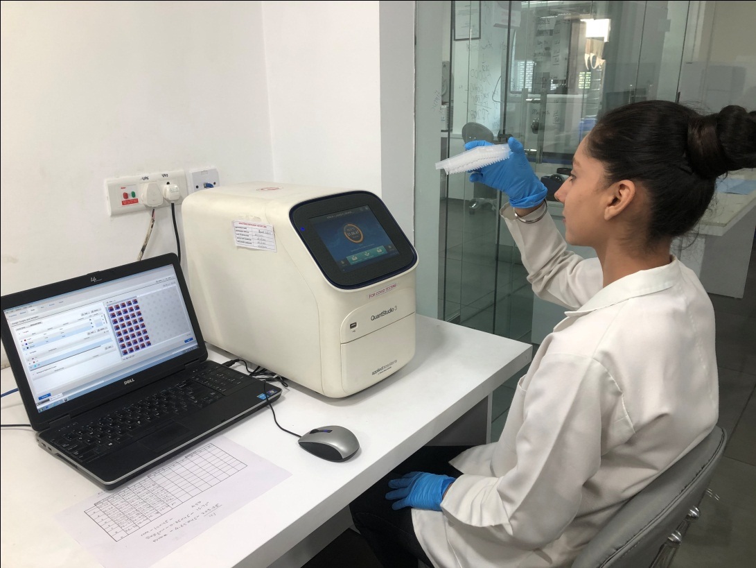
Real-time RT-PCR is currently the most utilized technique for detecting the coronavirus; however, several countries still, are in need of technical support in troubleshooting the use of this technique.
What is the genetic makeup of a virus?
Viruses are microscopic genetic materials that are encircled by another molecular envelope and usually they can be either a deoxyribonucleic acid or a ribonucleic acid. DNA is present in all organisms and it’s a two stranded molecule which holds the genetic code which is a blueprint of the way these organisms are made up of and how they develop. RNA on the other hand is single-stranded molecule which copies and it is transcribed and translated based on the genetic code to proteins which perform functions that maintain the life of the organism.
Viruses such as the SARS-CoV2 responsible for the coronavirus pandemic are comprised solely of ribonucleic acid thus suggesting that they have to infiltrate healthy cells in order to survive and multiply. Once the virus gets access into the cell, it uses its own genetic machinery to take over control of the cell and reprogram it into a virus making factory. As such for viruses like the corona virus to be detected within the body quickly using real-time RT-PCR.
Real-time COVID-19 RT-PCR detection process
The first step is to collect a sample from the possible elements of the body where the virus gathers such as the nasal cavity or throat. This is followed by treating the sample with a number of chemicals to remove substances such as proteins and fats to only remain with RNA which will be a mixture of the person’s genetic material and that of the coronavirus RNA. The reverse transcriptase enzyme is then used to reverse the RNA into deoxyribonucleic acid and then short fragments of DNA complementary to particular elements of the transcribed viral DNA are added to the reaction mixture. The fragments will then attach to the viral DNA sections if it is present. The added fragments also serve the purpose of building the DNA strands throughout the amplification process as well as adding labels to the strands which makes detection of the virus possible. The reaction mixture is then placed in the RT-PCR machine with specific temperature stages that heats and cools the mixture to initiate targeted chemical reactions to make new DNA strands of the virus. Usually, a standard real-time PCR is run for about 35 cycles which approximately amplifies about five billion new strands of the target viral DNA from a single copy present in the sample. The added marker labels also attach to the DNA as new strands get constructed and in the process release a fluorescent dye which is monitored by the computer and shown in real-time on the computer monitor. The quantity of the fluorescent within a sample is tracked once after every complete cycle and once the quantity of fluorescent crosses over a set threshold, it’s a confirmation that the target virus is present. The number of cycles it takes to succeed in this regard is also used as a measure of the severity of the infection and the fewer the cycles suggests that the viral infection is more.
Why real-time RT-PCR preferred in the detection of COVID-19?
The advantage of the real-time RT-PCR is that it is extremely sensitive and specific in delivering a reliable diagnosis in about two hours. Contrary to other available virus extraction methods available, the real-time RT-PCR is fast and has lower potential for errors or contamination and the entire process can be completed inside a closed tube hence the best method for the detection of the coronavirus. It is very important to observe past infections in order to understand the progression of the virus and since viruses are only present within the body of a particular time period which makes it difficult to use real-time PCR. Hence, other alternative strategies are required to monitor and track other past infections, particularly individuals who may have developed and spread the virus without showing any symptoms.
Conflict of interest:
There is no conflict of interest among the authors or anybody else and there has been no significant financial support for this work that could have influenced its outcome.
References
Guanine Cytosine rich GC rich regions represent a challenge.... https://www.coursehero.com/file/p31l0kf/GuanineCytosine-rich-GC-rich-regions-represent-a-challenge-in-standard-PCR/
How is the COVID-19 Virus Detected using Real Time RT-PCR. https://www.bignewsnetwork.com/news/264442648/how-is-the-covid-19-virus-detected-using-real-time-rt-pcr
IAEA Explains RT-PCR Virus Detection - News - Nuclear.... http://nuclearstreet.com/nuclear_power_industry_news/b/nuclear_power_news/archive/2020/04/01/iaea-explains-rt_2d00_pcr-virus-detection-040103
Reverse transcriptase is an enzyme that using an RNA.... https://www.coursehero.com/file/p2nmb50/Reverse-transcriptase-is-an-enzyme-that-using-an-RNA-transcript-can-make-a-DNA/
What are the differences between PCR, RT-PCR, qPCR, and RT.... https://www.enzolifesciences.com/science-center/technotes/2017/march/what-are-the-differences-between-pcr-rt-pcr-qpcr-and-rt-qpcr?/