A blood test using infrared spectroscopy can be used to diagnose two types of cancer, lymphoma and melanoma, according to a study led by Georgia State University.
Researchers used mid-infrared spectroscopy to analyze blood serum derived from experimental mice and differentiate mice with non-Hodgkin's lymphoma and subcutaneous melanoma from healthy mice and also between these two tumorous conditions. The mid-infrared spectral region of the electromagnetic spectrum is frequently used to characterize biological samples at the molecular level.
The findings, published in the journal Scientific Reports, suggest infrared spectroscopy can detect biochemical changes induced by non-Hodgkin's lymphoma, a solid tumorous condition of the immune system, and subcutaneous melanoma, a deadly form of skin cancer, and has diagnostic potential as a screening technique for these cancers.
Studies have found the incidence rates of cutaneous melanoma have increased in many regions and populations over the last decade, specifically 3 to 7 percent per year among fair-skinned populations. Also, non-Hodgkin's lymphoma accounts for 4.3 percent of new cancer cases in the United States. The available diagnostic regimen for both cancers, which includes tissue examination and biopsy, is time-consuming, invasive and costly, resulting in small compliance rates of eligible populations for cancer prescreening.
Developing a rapid and reliable prescreening strategy for melanoma and lymphoma is critical because early diagnosis and treatment of these malignancies improve the patients' chances of survival. Fourier Transform Infrared (FTIR) spectroscopy in Attenuated Total Reflection (ATR) sampling mode provides high-quality results with better reproducibility compared to other vibrational spectroscopy. It has attracted scientists' attention for its rapid and reliable detection of various health conditions using body fluid samples.
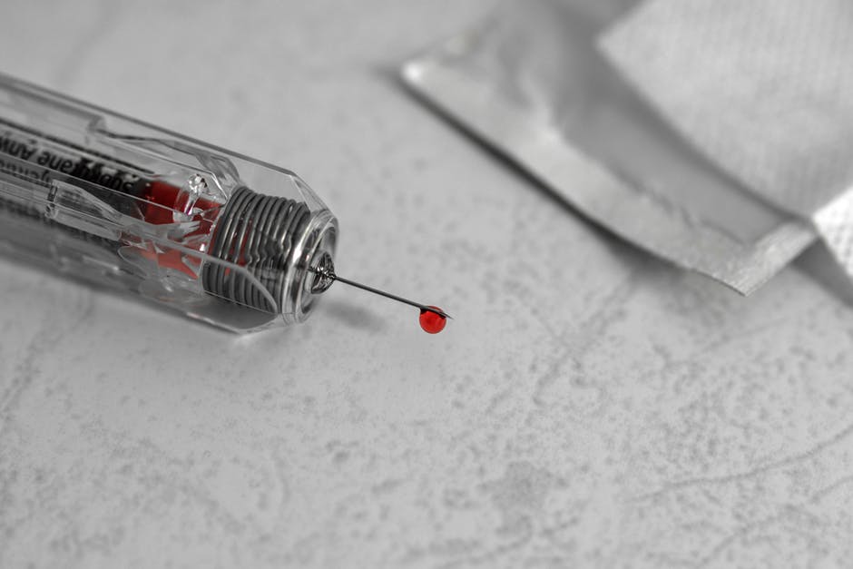
In this recent study, the researchers used mice with lymphoma and melanoma cancers. Blood serum droplets extracted from cancerous mice and control mice were placed on an ATR crystal of the FTIR instrument. Incident infrared beams were absorbed and reflected by the serum, creating a wave that was recorded and used to produce an absorbance curve with peaks that identified the presence of certain biomarkers in the sample.
The researchers compared the absorbance curves from the control and tumorous mice and assessed biochemical changes induced by non-Hodgkin's lymphoma and subcutaneous melanoma in the serum samples obtained from Dr. Yuan Liu's research lab in Georgia State's Department of Biology.
The study found remarkable differences between the ATR-FTIR spectra of serum samples from tumor-bearing mice with melanoma and non-Hodgkin's lymphoma and healthy, control mice.
The findings are applicable to humans because mice and humans have some biomarkers and chemicals in common, Perera said. In previous studies on colitis, Perera and his colleagues identified specific chemicals that changed in humans and mice when colitis was present.
Using the data collected on the biomarkers for lymphoma and melanoma, the researchers can develop detectors for these particular absorbance peaks, which doctors could use to test patients' blood samples for these cancers.