James Clerk Maxwell during 1860s gave the theory that light is an electromagnetic wave having electric (E) and magnetic (B) field components and interaction of light with matter not only experience phenomena’s like reflection, refraction, absorption, transmission etc. but light waves or EM waves interacting with electrons in the matter also exert force on the matter leading towards the generation of optical pressure or light pressure or radiation pressure.
Optical pressure
Radiation pressure is the force per unit area on an object due to change in light momentum. The light momentum of a single photon is:

The change in momentum can be calculated by the difference in momentum flux between entering and leaving an object

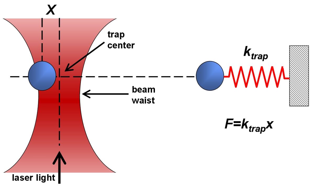
The force exerted by light or the optical pressure experienced by a macro object is almost negligible but in case of a micro or nano size object even the small amount of pressure cannot be ignored. Further, using a laser light (which is more intense, coherent, and unidirectional) on a microscopic particle will realize more the situation of optical pressure development. Force develops when rays of light act on objects with different index of refraction compared to the surrounding media. The particle gets push towards the highest light intensity. However there are also reflections pushing the bead forward.
With the development of laser in 1960s, scientists started working in the field of acceleration and trapping of particles by optical pressure. Micron-sized particles have been accelerated and trapped in stable optical potential wells using only the force of radiation pressure from a continuous laser. It is hypothesized that similar accelerations and trapping are possible with atoms and molecules using laser light tuned to specific optical transitions. A particle can get sucked into the focus of a laser bundle and be stably trapped. Thus, highly focused laser beam acts as a three-dimensional potential minimum. Therefore, it takes force to dislodge a bead out of the laser focus. The research manipulating the control of particles at micro level with laser light was recognized and Physics Nobel prize-1997 was awarded jointly to Steven Chu, Claude Cohen-Tannoudji and William D. Phillips for their developments of methods to cool and trap atoms with laser light. Researchers have developed methods of cooling and trapping atoms by using laser light and the research is helping to study fundamental phenomena and measure important physical quantities with unprecedented precision.
Optical tweezers
Optical tweezers developed in 1980s use forces of laser radiation pressure to trap small objects and today are known as the useful micro-manipulation laser tool. Optical tweezers use light to manipulate microscopic objects as small as a single atom. The radiation pressure from a focused laser beam is able to trap small particles. The most basic form of an optical trap is diagramed below. A laser beam is focused by a high-quality microscope objective to a spot in the specimen plane. This spot creates an "optical trap" which is able to hold a small particle at its center. The forces felt by this particle consist of the light scattering and gradient forces due to the interaction of the particle with the light. Most frequently, optical tweezers are built by modifying a standard optical microscope. These instruments have evolved from simple tools to manipulate micron-sized objects to sophisticated devices under computer control that can measure displacements and forces with high precision and accuracy.

In practice, optical tweezers are very expensive, custom-built instruments. These instruments usually start with a commercial optical microscope but add extensive modifications. In addition, the capability to couple multiple lasers into the microscope poses another challenge. High-power infrared laser beams are often used to achieve high trapping stiffness with minimal photodamage to biological samples. Precise steering of the optical trap is accomplished with lenses, mirrors, and acousto/electro-optical devices that can be controlled via computer. The diagram below is meant to give an idea of the number of elements in such a system. In short, these are very complicated instruments that require a working knowledge of microscopy, optics, and laser techniques.
The earlier trap was utilized two sources of laser beams or the combination of one laser beam and gravitational force to achieve the stability of the trap by Ashkins 1980. In 1986, Ashkin et al. has published the first single beam optical tweezers. Now, the technique has been improved and customized to suit the researcher’s need. This year’s (2018) Nobel Prize in physics has gone to three scientists for creating what has been described as “tools made of light”. Prof. Arthur Ashkin (one of the Nobel Laureate of Physics2-18) is credited with having invented what is famously known as “optical tweezers”. Actually a technology rather than a physical instrument, these “tweezers” are widely used for isolating and examining very small particles, such as individual atoms, DNA strands, or biological cells.
Exotic applications
One of the major goals of modern day science is to describe and characterize the world as it exists on the scales that are far beyond the scope of our own sense as the information can be used in various ways to benefit our lives. Manipulating small objects is one of the quests in finding the information beneficial to our own. The challenge was to find a precise, easy to manipulate tool, and it must provide minimal damage to the studied object.
The practical applications of optical tweezers are endless. Optical tweezers have been used to trap dielectric spheres, viruses, bacteria, living cells, organelles, small metal particles, and even strands of DNA. Two of the main uses for optical traps have been the study of molecular motors and the physical properties of DNA. In both areas, a biological specimen is biochemically attached to a micron-sized glass or polystyrene bead that is then trapped. By attaching a single molecular motor (such as kinesin, myosin, RNA polymerase etc.) to such a bead, researchers have been able to probe motor properties such as: whether the motor take individual steps, the step size, force which the motor can produce. Similarly, by attaching the beads to the ends of single pieces of DNA, experiments have measured the elasticity of the DNA, as well as the forces under which the DNA breaks or undergoes a phase transition. Applications include:
- pull or displace microscopic particles
- altering of larger structures (such as cell membranes)
- measure microscopically small forces like: • studying the strength of biological materials such as cells, membranes, proteins or DNA
- confinement and organization (e.g. for cell sorting)
- detection of force generation in molecular motors such as kinesin (the protein responsible for pulling apart chromosomes during cell division) or RNA polymerase
- elucidation of the microscopic properties of complex solutions (for example: polymer solution)
- tracking of movement (e.g. of bacteria)