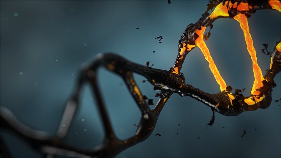
It's DNA, but not as we know it.
In a world first, Australian researchers have identified a new DNA structure called the i-motif inside cells. A twisted 'knot' of DNA, the i-motif has never before been directly seen inside living cells.
The new findings, from the Garvan Institute of Medical Research, are published today in the leading journal Nature Chemistry.
Deep inside the cells in our body lies our DNA. The information in the DNA code all 6 billion A, C, G and T letters provides precise instructions for how our bodies are built, and how they work.
The iconic 'double helix' shape of DNA has captured the public imagination since 1953, when James Watson and Francis Crick famously uncovered the structure of DNA. However, it's now known that short stretches of DNA can exist in other shapes, in the laboratory at least and scientists suspect that these different shapes might play an important role in how and when the DNA code is 'read'.
The new shape looks entirely different to the double-stranded DNA double helix.
When most of us think of DNA, we think of the double helix. This new research reminds us that totally different DNA structures exist -- and could well be important for our cells.
Although researchers have seen the i-motif before and have studied it in detail, it has only been witnessed in vitro -- that is, under artificial conditions in the laboratory, and not inside cells.
In fact, scientists in the field have debated whether i-motif 'knots' would exist at all inside living things -- a question that is resolved by the new findings.
To detect the i-motifs inside cells, the researchers developed a precise new tool a fragment of an antibody molecule -- that could specifically recognise and attach to i-motifs with a very high affinity. Until now, the lack of an antibody that is specific for i-motifs has severely hampered the understanding of their role.
Crucially, the antibody fragment didn't detect DNA in helical form, nor did it recognise 'G-quadruplex structures' (a structurally similar four-stranded DNA arrangement).
With the new tool, researchers uncovered the location of 'i-motifs' in a range of human cell lines. Using fluorescence techniques to pinpoint where the i-motifs were located, they identified numerous spots of green within the nucleus, which indicate the position of i-motifs.
