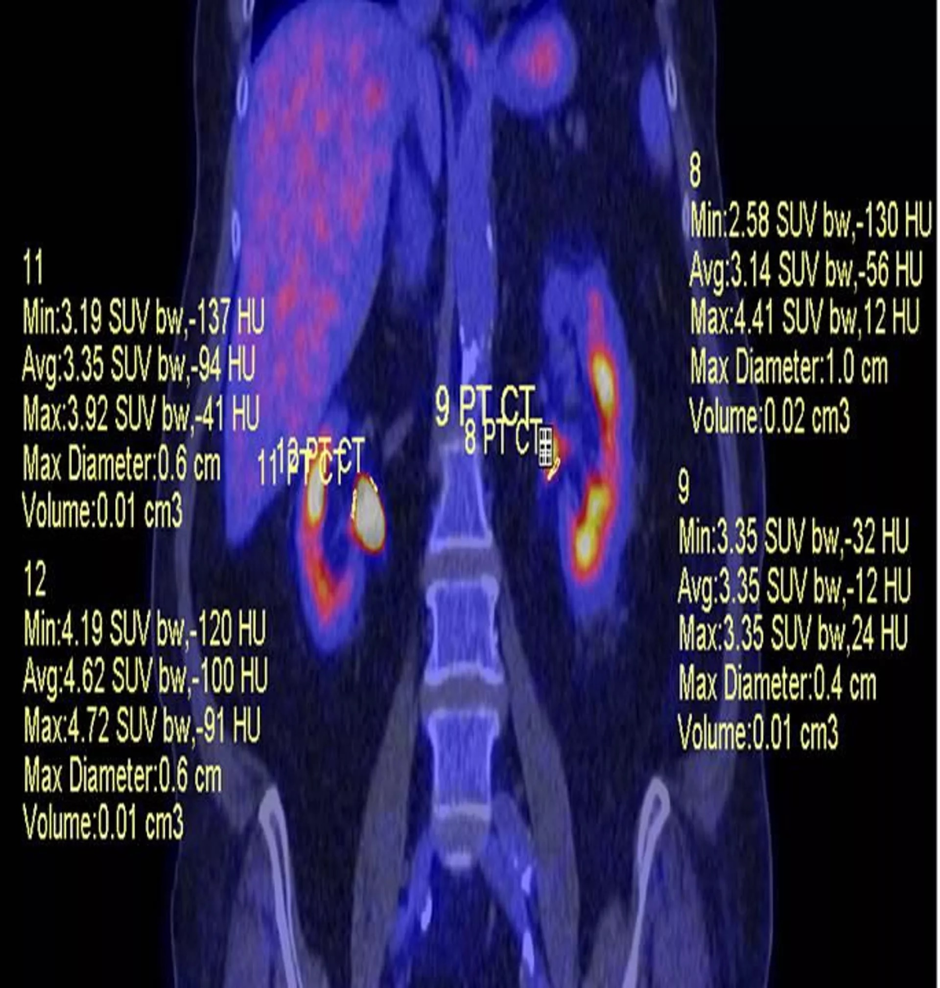
Scientists from the Tyumen State Medical University of the Russian Ministry of Health (TyumSMU) believe that early detection of chronic kidney disease is possible using combined positron emission tomography and computed tomography (PET /CT).
Chronic kidney disease (CKD) can develop as a result of several diseases such as pyelonephritis, urolithiasis, polycystic disease, and others, the university's scientists explained. This diagnosis leads to renal failure and cardiovascular complications.
CKD is one of the "silent" diseases. It is considered problematic to detect it before the late stages. When dead cells are found in the patient's tests, the process of tissue destruction is irreversible, and medicine can only slow it down. The disease is diagnosed in 10-15% of the adult population of Russia.
Researchers at TyumSMU have found that PET/CT can detect this and other dangerous kidney diseases in the early stages. At a time when there are no complaints or changes in urine tests, but the disease is already forming at the molecular and cellular level, which is not accessible by any of the existing diagnostic methods.
“PET/CT is a high-tech imaging of carbohydrate and lipid metabolism in various organs, resulting in a real-time view of the molecular and cellular viability of tissue. Previously, this method was used only in oncology diagnostics. We found out that it can be used to detect kidney disease at an early stage. The patient is injected with labeled glucose molecules, observing their fixation by kidney cellular structures, we can see and calculate the viability of the organ, as well as trace regions of decreased metabolic activity,” said Professor Boris Berdichevsky at TyumSMU’s Department of Surgery and Urology with Endoscopy Course.
Tumors "love" glucose and light up brightly on the radiologist's screen, the specialist explained. Meanwhile, normal organs cannot ensure the normal vitality of the human body without glucose, either. Previously, doctors did not pay attention to the difference between its capture by healthy cells and those that are already prone to disease. The latter are at the stage of "tissue stunning," and if the problem is not identified at this stage, the organ slowly dies.
"The cell decreases its glucose intake at the onset of any disease that affects the tissues, it is as if its ‘appetite’ drops. When the amount of absorbed carbohydrate decreases by 10%, there is no obvious danger, but if the figure exceeds 30%, it means that the cells are already "stunned" and it is time to select an appropriate therapy," Professor Berdichevsky continued.
The scientist believes that PET/CT may eventually replace conventional fluorography in the course of medical examinations of the country's population.
According to the findings of the scientists at Tyumen Medical University, positron emission computed tomography can be used to detect metabolic disorders of any organ long before their clinical manifestations appear. The results of their study were published in the Urology journal.
The university's scientists believe that detailed studies of its effectiveness in detecting various diseases can accelerate the introduction of the method in preventive diagnostics.
